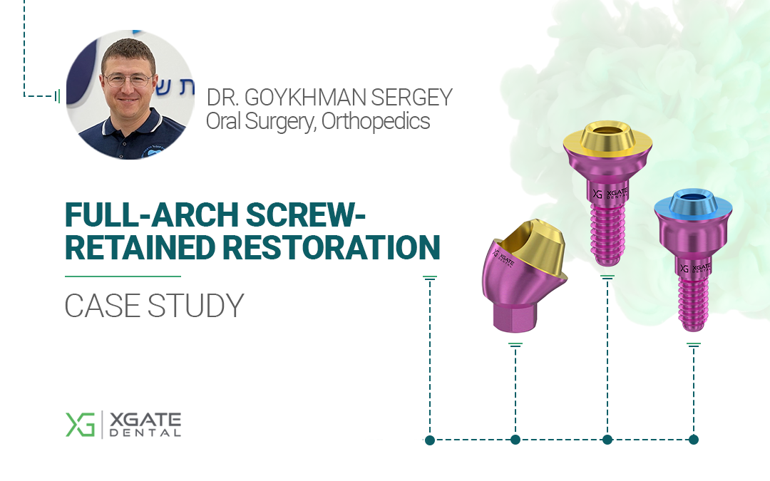
We present to your attention the full-arch restoration clinical case that showcases the use of 4-6 implants and superstructures from different brands, highlighting the advantage of interface standardization. This interesting and high-quality case analysis will be valuable for both new and experienced specialists.
First, let’s get acquainted with the doctor:

Specialization: Oral Surgery, Orthopedics, Restorative Dentistry,
Soft Tissue Management
Experience: 16 years
Brief Patient Information
The patient is a 65-year-old woman presenting with discomfort from her maxillary denture. She is systemically healthy, with no chronic diseases. She had previously worn removable maxillary dentures. The maxilla was completely edentulous, while the mandible was partially edentulous, with several teeth remaining in the anterior region. Implant-supported bridges were present in the posterior regions of the mandible.

The decision was made to extract the remaining mandibular teeth, place implants, and fabricate a new implant-supported prosthesis.
For the maxilla, it was planned to place six implants with a restoration according to an All-on-6 protocol.
Treatment Protocol
Diagnostic stage:
- CBCT (Cone Beam Computed Tomography);
- Intraoral scanning of both arches.

Digital CAD/CAM technologies were extensively utilized for this patient, making screw retention an ideal choice. For 3D modeling, virtual libraries of implants, multi-units, screws, and other components are available. These virtual components can be easily placed onto a 3D model of the jaws to verify their fit. The virtual prosthesis model can then be positioned onto these components, allowing for adjustments to implant position or angulation, or selection of different abutments, if needed.
Treatment planning stage
1. Manufacturing of a surgical guide for the placement of 6 maxillary and 4 mandibular implants. Maxilla: An All-on-6 approach was planned, with the most distal implants placed at a 30° angle to bypass the maxillary sinus and engage sufficient preserved bone volume. Mandible: Implant positions were calculated to ensure even load distribution and integrate seamlessly with previously placed implants.

2. A surgical guide for precise implant positioning was designed and manufactured in a virtual environment.
MIS implants were selected. The interface chosen was a standard hexagon. Implant diameters were 4.2 mm for the maxillary posterior regions and 3.75 mm for the anterior regions of both the maxilla and mandible.
Surgical stage of treatment
1. Extraction of the anterior mandibular teeth was performed simultaneously with the placement of four 3.75 mm diameter implants. Teeth were extracted atraumatically, and the existing dentures were preserved.

The implants were placed close to the original tooth positions:
Tooth #32 – 1 mm abutment height
Tooth #33 – 2 mm abutment height
Tooth #42 – 1 mm abutment height
Tooth #44 – 1 mm abutment height
All implants were placed with a torque greater than 35–45 Ncm, allowing for immediate loading. Abutments were torqued to 30 Ncm, which is the standard protocol.
2. For prosthetics, we primarily selected XGate V-type multi-unit abutments, which feature a smaller cone but an increased contact area (10 mm²) between the abutment platform and the prosthesis sleeve, compared to the 6 mm² of the D-type standard. This configuration allows for greater bulk of the prosthesis body at the interface with the abutment.

Six implants were placed in the maxilla at the following positions:
#11 – 1 mm V-type abutment
#13 – 2 mm V-type abutment
#16 – 1 mm D-type angled abutment (30°), to compensate for implant angulation relative to the occlusal plane.
#21 – 1 mm V-type abutment
#23 – 2 mm V-type abutment
#26 – 1 mm D-type angled abutment (30°)
V-Type & D-Type Multi-Units

1mm straight MUA
V-type 5201.1001

2mm straight MUA
V-type 5201.1002

1 mm 30° MUA
D-type 5101.1031

Stage of prosthetics and healing
After implant placement, scan bodies were installed to accurately record the abutment positions. This procedure was performed for both maxillary and mandibular arches, a necessary step for the fabrication of both temporary and definitive prostheses.


For the mandible, existing bridges were removed, and new abutments and scan bodies were placed on the previously installed implants.

Following scanning, healing caps were placed, and soft tissues were sutured.

After implant placement, the patient was discharged. Temporary PMMA prostheses were fabricated the following day.
The next day, healing caps were removed, and the temporary complete maxillary denture was installed.


Following healing and osseointegration, the patient received permanent zirconia dioxide restorations
In the maxilla, two multi-unit abutments were replaced because the healed gingival tissue was higher than at the time of implant placement. Consequently, the 1 mm V-type multi-unit abutments were replaced with 3 mm V-type abutments, as shown in the following photo.


Two bridges were fabricated for the maxilla, separated at the central incisors, to minimize internal stress and facilitate seating.
Mandible: The restoration was divided into three bridges, accounting for the previously placed implants.


XGate Dental Products
This clinical case demonstrates the use of both XGate V-type and D-type abutment systems. Use the jaw filter below to view specific components for the maxilla or mandible.
Download Resources
Get the complete case study and explore our product catalog
Case Study • PDF • 1.6 MB
📥 Download Case StudyProduct Catalog • Online
🔍 View Product CatalogWe hope you found this clinical case interesting.If you have any questions about the characteristics and delivery of XGate Dental products, please contact us in any convenient way.
E-mail: [email protected]
350 W Passaic
St Rochelle Park, NJ 07662
United States
XGate Dental Group GmbH
Falkensteiner Straße 77, 60322
Frankfurt am Main
Germany
Disclaimer: Any medical or scientific information provided in connection with the content presented here makes no claim to completeness and the topicality, accuracy and balance of such information provided is not guaranteed. The information provided by XGATE Dental Group GmbH does not constitute medical advice or recommendation and is in no way a substitute for professional advice from a physician, dentist or other healthcare professional and must not be used as a basis for diagnosis or for selecting, starting, changing or stopping medical treatment.
Physicians, dentists and other healthcare professionals are solely responsible for the individual medical assessment of each case and for their medical decisions, selection and application of diagnostic methods, medical protocols, treatments and products.
XGATE Dental Group GmbH does not accept any liability for any inconvenience or damage resulting from the use of the content and information presented here. Products or treatments shown may not be available in all countries and different information may apply in different countries. For country-specific information please refer to our customer service or a distributor or partner of XGATE Dental Group GmbH in your region.
