Contents
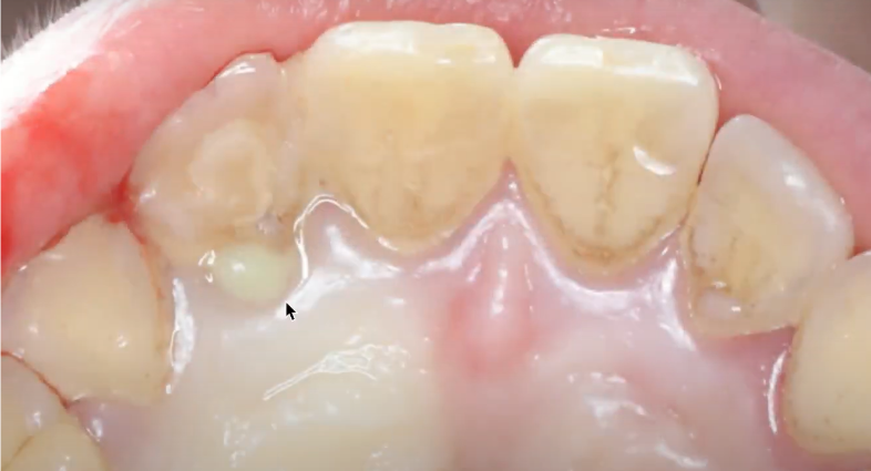
We concluded the previous part with a clinical case of immediate implantation using the Socket Shield technique, highlighting it as one of the most effective methods for preserving the gingiva and the buccal plate, especially in the anterior maxilla. Let’s briefly review that case. The patient presented with pain and mobility of tooth #12 (FDI notation). The cause was a traumatic luxation (dislocation), and a year and a half of unsuccessful endodontic treatment in an attempt to regain periodontal health.

Pus discharge from the palatal side of the problem tooth (12) YouTube / Dr. Kamil Khabiev / Dental Guru Academy
Pus discharge from the palatal side of the problem tooth (12) YouTube / Dr. Kamil Khabiev / Dental Guru Academy
The second picture illustrates the prepared implant site, where a fragment of the tooth root remains on the buccal side, acting as a shield to protect the gingiva and maintain the height of the buccal bone.

Buccal wall (shield) ready for further manipulations: color and condition of completely healthy tissue YouTube / Dr. Kamil Khabiev / Dental Guru Academy
The third photograph displays the final result, a few months after the definitive crown was placed. Without knowing we’re focusing on tooth #12, it’s difficult to discern any differences in the gingival margin.

Definitive crown on tooth #12 – excellent functional and aesthetic outcome YouTube / Dr. Kamil Khabiev / Dental Guru Academy
While a successful clinical case is encouraging, it’s not conclusive. However, a significant number of similar patients exist, and the data is continuously updated and analyzed. One particularly relevant study is: “The Root Membrane Technique: Human Histologic Evidence after Five Years of Function”. by Dr. Miltiadis E. Mitsias and his team. In this study, a volunteer (a 68-year-old male) had an implant and surrounding tissues removed after 5 years of function. The implant had been placed using the Root Membrane (RM) technique; Socket Shield is a variation of Root Membrane (RM).
The researchers conducted a histological analysis of the samples and confirmed that root fragment preservation techniques during implant placement in the anterior region effectively maintain the buccal plate. Here is a direct quote from the study:
“The buccal bone plate was preserved without any resorption; a healthy periodontal ligament was confirmed. The implant demonstrated osseointegration with a high percentage of bone-to-implant contact (BIC = 76.2%). Regarding the space between the RM and the implant, the apical and medial thirds were filled with compact mature bone; the coronal third was colonized by non-infiltrated connective tissue.”
Here are images of the histological sections:

Trabecular, mature bone at the interface of the implant was observed. The bone was present between the implant and the root. The root membrane and the buccal bone plate appeared intact without any signs of resorption. Acid fuchsin-toluidine blue 12x. YouTube / Dr. Kamil Khabiev / Dental Guru Academy

Compact bone in the medial thirds and apical portion of the implant were evident. No gaps were present at the interface. Acid fuchsin-toluidine blue 40x. YouTube / Dr. Kamil Khabiev / Dental Guru Academy

In the coronal portion, between the root and the implant, connective tissue without inflammatory infiltrate was present. Acid fuchsin-toluidine blue 40x. YouTube / Dr. Kamil Khabiev / Dental Guru Academy

In the apical portion of the root, it was observed that the cementum migrated from the residual root to the implant surface. Acid fuchsin-toluidine blue 40x. YouTube / Dr. Kamil Khabiev / Dental Guru Academyм
Assessing Primary Implant Stability
Primary insertion torque is an unreliable indicator of implant stability; specialized instruments are needed. Many clinicians utilize devices like the Osstell ISQ or similar tools to determine the actual stability of implants. In the following illustration, the stability of an implant at the site of the second incisor is being checked. According to the instrument readings, immediate loading is appropriate in this case.

Mega-ISQ readings during primary implant stability assessment YouTube / Dr. Kamil Khabiev / Dental Guru Academy
These devices operate on the principle of Resonance Frequency Analysis (RFA), which is a precise method. This method is based on measuring the vibration frequency of the implant, which is correlated to its stability in the bone.
Here’s a review of the operating principle:
- A small magnetic peg or abutment (disposable or reusable) is attached to the implant. Stability is measured in several planes, as illustrated:

Measuring Primary Implant Stability Using Resonance Frequency Analysis (RFA) YouTube / Dr. Kamil Khabiev / Dental Guru Academy
- The device emits vibrations and measures the resonant frequency.
- The frequency is converted to a numerical value – ISQ (Implant Stability Quotient), indicating the level of implant stability:
- Low ISQ: May indicate insufficient stability.
- High ISQ (greater than 65): Indicates good implant osseointegration.

Primary Implant Stability Diagram Based on ISQ Value YouTube / Dr. Kamil Khabiev / Dental Guru Academy
But perhaps not everyone knows about the new generation of compact devices that utilize the same principle. The photo below shows an example. These are often more convenient for immediate implant placement procedures, allowing the clinician to quickly assess whether immediate loading is suitable, or if a delayed loading protocol is necessary.

A device for measuring the primary stability of an implant, utilizing the same principle of resonance frequency analysis (RFA) YouTube / Dr. Kamil Khabiev / Dental Guru Academy
The device in the image shows an ISQ of 77, a very favorable result. This ISQ score allows for immediate loading in the anterior region or early loading after 1-2 months, even if a delayed loading protocol was initially planned.
Factors Influencing the Success of Immediate Implantation
- Bone type. Denser bone provides more points of contact between the bone and the implant surface. As shown below, in D4 bone, only a small fraction of the implant surface is in contact.

The difference in implant contact area in varying bone densities (in D2 bone, the contact area of titanium with bone is much larger) YouTube / Dr. Kamil Khabiev / Dental Guru Academy
- The implant surface: The roughness, topography, and cleanliness of the implant surface are critical. SLA surface treatment, and enhanced modifications, have demonstrated favorable outcomes. Osteoblasts readily attach to such a surface, leading to faster osseointegration and improved secondary stability, enabling earlier placement of definitive restorations.
Challenges and Solutions for Immediate Implant Placement in the Chewing Areas
In the molar region, bone grafting is generally avoided. Implants are typically placed in the inter-radicular septum, and a healing abutment is immediately placed, as shown in this image.

Immediate implant placement following molar extraction YouTube / Dr. Kamil Khabiev / Dental Guru Academy
This is standard practice. However, challenges such as gingival cuff leakage, as shown below, sometimes arise.

Poorly formed soft tissue attachment around the healing cap YouTube / Dr. Kamil Khabiev / Dental Guru Academy
This issue is not as rare as we’d like, and often requires suturing the soft tissues tightly before re-installing the healing cap. In some cases, impaired healing can lead to implant failure.
This often stems from improper selection of the healing cap. In the image above, the abutment is too narrow, leading to wound dehiscence and loss of the blood clot. Large posterior teeth have irregular shapes, making it difficult to select a standard round healing abutment. A custom healing cap can address this.
This well-established procedure utilizes a temporary abutment and flowable composite to ideally fill the soft tissue defect following atraumatic extraction. Preserving the gingiva during extraction is crucial.

Fabrication of a custom healing cap (left) and the healing outcome three days after placement (right) YouTube / Dr. Kamil Khabiev / Dental Guru Academy
Here’s a summary of the procedure: Install an appropriately sized abutment in the implant. Apply flowable composite to fill the extraction socket, embedding the abutment within the composite. Polymerize the composite, then remove it together with the abutment. Grind and refine the composite to its ideal form. Finally, re-install the custom healing cap and leave it in place until healing is complete. This preserves the emergence profile of the extracted tooth.

Well-formed gingiva prepared for restoration YouTube / Dr. Kamil Khabiev / Dental Guru Academy
Here’s what the restoration looks like in this clinical case:

Final stage: custom abutment for cement-retained restoration (left) and definitive crown (right) YouTube / Dr. Kamil Khabiev / Dental Guru Academy
The soft tissues were fully preserved, and the soft tissue connection to the abutment is hermetic. With a two-stage approach, guided bone regeneration or soft tissue grafting would likely have been necessary. Immediate implantation is more comfortable for the patient, involving fewer surgical procedures, less pain, and a shorter healing period.
Immediate Implantation in Full-Arch Restorations
Let’s consider a challenging clinical case involving a patient over 60 years of age. The image below reveals a large cyst associated with several teeth in the anterior segment of the mandible. The surgeon opted not to place implants in this section.

A snapshot of the patient’s initial situation – a large cyst rendering the anterior mandible unsuitable for implant placement YouTube / Dr. Kamil Khabiev / Dental Guru Academy
A failing removable partial denture (RPD) and ill-fitting PFM restorations had compromised the quality of the bone. The remaining teeth cannot be salvaged for future restoration, as is evident in the image below.

Severely damaged teeth resulting from RPD wear YouTube / Dr. Kamil Khabiev / Dental Guru Academy
The RPD design is flawed; the distal extensions are supported only by soft tissue, concentrating the load on the anterior teeth. The distal extensions act as levers during chewing, leading to this situation in less than three years.

RPD after three years of use YouTube / Dr. Kamil Khabiev / Dental Guru Academy
The aesthetics of this type of restoration are also lacking. Dark shadows are visible at the margins of the restoration and unhealthy gingiva.

Patient with a failing RPD: poor appearance and compromised function YouTube / Dr. Kamil Khabiev / Dental Guru Academy
Given the complexities of this case, ridge reduction was necessary to facilitate a functional prosthesis. Esthetics remained a secondary concern.
To maintain functionality in the future, an impression was taken to capture the centric relation of the jaws. The definitive restoration was then fabricated based on this impression, ensuring compatibility with the opposing dentition. The old crowns were placed in the RPD and the patient was asked to bite down on a registration material (e.g., vinyl polysiloxane).

Making a cast to register the position of the jaws and the position of the dental arches YouTube / Dr. Kamil Khabiev / Dental Guru Academy
Creating a full cast, precisely capturing the palatal portion, is essential in these situations. This “palatal stop” provides a baseline for creating the restoration after altering the alveolar ridge.

Full cast of the upper jaw with the palatal part, which will help in the production of restoration YouTube / Dr. Kamil Khabiev / Dental Guru Academy
We then proceeded with ridge reduction. Alveoloplasty was performed to improve esthetics by ensuring the gingiva doesn’t display when smiling. The image below shows alveoloplasty (top), and assessment of primary implant stability with a Penguin device (bottom).

The process of bone ridge reduction with immediate implant placement, in the lower image the primary stability of the implant is checked by compact resonance frequency analysis (RFA) device YouTube / Dr. Kamil Khabiev / Dental Guru Academy
All implants exhibited excellent primary stability, despite the patient’s poor bone density, as confirmed by the instrument readings. This allowed immediate loading and for the patient to leave the clinic with teeth on the day of surgery.

Instrument readings to determine the primary stability of placed implants: all implants are placed with very good indicators, which allow immediate loading of the implants YouTube / Dr. Kamil Khabiev / Dental Guru Academy
Next, the bite registration, obtained before extractions, was used to determine the jaw relationship and create a fully functional jaw model.

Bite registration after extraction and implant placement YouTube / Dr. Kamil Khabiev / Dental Guru Academy
The bite registration was covered with bite registration paste and the postoperative relief of the maxilla was recorded. The model created from this bite is crucial for the dental technician during the creation of a full denture.

Postoperative maxillary relief recording for the laboratory YouTube / Dr. Kamil Khabiev / Dental Guru Academy
The next step was taking an impression. Because the implants were placed at varying angles, the impression copings (transfers) were splinted to ensure mechanical rigidity and prevent micro-movements when the impression was made.

Combining transfers into a single structure to provide mechanical strength before taking an impression YouTube / Dr. Kamil Khabiev / Dental Guru Academy
The patient was then prepared for placement of a temporary restoration. The image below shows the condition of the maxilla two days post-surgery. The ridge contour looks good, and the reduction was successful.

Condition of soft tissue before placement of the temporary prosthesis YouTube / Dr. Kamil Khabiev / Dental Guru Academy
The temporary restoration looked quite acceptable and was seated directly over the sutures, as is standard in such procedures.

Temporary restoration in place for healing and osseointegration YouTube / Dr. Kamil Khabiev / Dental Guru Academy
This shows the patient’s smile four months after the procedure, with a definitive restoration that mimics the appearance of gingiva. The soft tissues under the prosthesis are healthy and healing is progressing well. The patient was very pleased with the results, which included:
- A single comprehensive surgical procedure including ridge reduction and implant placement.
- A short healing period (averaging 4 months).

Permanent restoration with excellent aesthetic results YouTube / Dr. Kamil Khabiev / Dental Guru Academy
Digital Technologies in the Protocol of Immediate Implantation
Digital technologies have the potential to significantly reduce the time commitment for both the patient and the dentist, allowing the patient to leave the office with a temporary prosthesis just hours after the procedure. This can be accomplished by replacing traditional impressions, which many patients find uncomfortable, with two digital files: The first is a DICOM file from a cone-beam computed tomography (CBCT) scan.

Cone Beam CT Scan of the Jaws (image from Shutterstock)
The second is an STL file from an intraoral scanner, which captures the condition of the remaining teeth and surrounding soft tissues.

Intraoral scanner for taking virtual impressions of jaws (image from Shutterstock)
Software can merge the STL and DICOM files into a single working 3D model of the patient’s jaws. The digital model can be used to:
- Plan the exact location of the implants.

Planning the location of the implants on a digital 3D model of the jaws (image from Shutterstock)
- Design and fabricate custom abutments, temporary restorations, and surgical guides.
These steps can be performed without the patient being present, requiring only a tomogram and an intraoral scan. This allows the dentist to have a temporary prosthesis, surgical guide, and, if needed, custom abutments available on the day of the surgery.
Software packages such as Exocad DentalCAD or 3Shape Dental System are commonly used. Some manufacturers, like Nobel Biocare and R2Gate, have created specialized software.
In the following example, we used R2 Gate software.
The patient was missing a significant number of teeth in the anterior mandible. The bone density was determined to be type 2-3.

Initial situation – significant loss of teeth in the lower jaw YouTube / Dr. Kamil Khabiev / Dental Guru Academy
The first stage of treatment involves:
- Placement of implants in positions 32, 41, 43, 46, and 47 (FDI).
- Design and fabrication of a temporary prosthesis with temporary abutments.
- Securing the temporary prosthesis.
The most challenging and difficult processes were simplified and radically altered by digital technology.
First, a wax template on a rigid base was used to determine the bite. The patient bites the bite roller, fixing the central relationship of the jaws.

Wax bite roller for fixing the central relationship of the jaws YouTube / Dr. Kamil Khabiev / Dental Guru Academy
Next, the wax roller with the imprint of the upper teeth, the teeth, and soft tissues are treated with a special matte spray to eliminate glare and transparency of the surfaces. This significantly increases the scan’s accuracy.

Model of a patient’s jaw with a wax bite block YouTube / Dr. Kamil Khabiev / Dental Guru Academy
An operation plan is created on a 3D model. The exact position of the implants is determined and a prosthesis is designed based on the upper jaw and antagonist teeth. The illustration shows how a prosthesis can be created.

Modeling the optimal locations of implants and the prosthesis that will rest on these implants YouTube / Dr. Kamil Khabiev / Dental Guru Academy
Custom abutments for the retention of the temporary prosthesis are made in digital format. The patient does not need to come into the clinic for impressions.
A combination of computer tomography and optical scanning allows the clinician to see both the state of the bone and the level of the gums. This allowed the wax roller to be used to fix the gingival height.

Part of the workflow: modeling the prosthesis taking into account the gingival height and correcting the location of the implants YouTube / Dr. Kamil Khabiev / Dental Guru Academy
The abutments can be seen from all angles and the clinician can ‘remove’ and ‘return’ bone and soft tissue from the image.

Overlay image of implants with virtual scan markers and prosthesis YouTube / Dr. Kamil Khabiev / Dental Guru Academy
After 3D modeling has been completed, all the data is sent to the laboratory, where the following are manufactured:
- custom abutments (temporary)
- surgical template
- temporary restoration

The temporary abutment and restoration are made and are kept by the doctor before the operation YouTube / Dr. Kamil Khabiev / Dental Guru Academy
The abutments are milled from polymethyl methacrylate. The abutments are light, but still, hold the temporary restoration. If an unforeseen situation occurs, the abutment will break before damage to the implant.

The difference in weight between a polymer and titanium abutment of the same shape YouTube / Dr. Kamil Khabiev / Dental Guru Academy
A key advantage of R2 Gate and similar software is the high precision in manufacturing and the excellent passive fit of the restoration. This is because the software considers not only the condition of all tissues and the patient’s anatomy but also precisely plans the implant’s fit, down to the position of the hexagon of the internal interface. This precision is critical; otherwise, it’s impossible to install the individual abutments in the same position as designed in the virtual model. A surgical template with precisely placed slots, used in combination with a special implant driver, enables this accurate positioning of the implant along its edges. Now, let’s review the photo protocol of the procedure. The first image shows the initial condition of the patient’s jaw. Before installing the surgical template, a longitudinal incision is made (as seen in the second photo). The soft tissues are then reflected to the buccal and lingual aspects, providing access to the alveolar ridge for proper placement of the surgical template


Initial state of the edentulous area of the jaw (upper image) and the first longitudinal incision for access to the alveolar ridge YouTube / Dr. Kamil Khabiev / Dental Guru Academy
Of particular note is the surgical template, which, while partially supported by the patient’s remaining tooth, relies primarily on bone-based retention. However, the most significant feature is the presence of slots on the outer aspect of the guide bushings.

A digitally designed and 3D printed individual surgical template for precise positioning of implants including the position of the internal interface edgesYouTube / Dr. Kamil Khabiev / Dental Guru Academy
These slots are crucial for precisely positioning the hexagon within the implant interface. The goal is to install the implants exactly as planned in the virtual model, controlling both depth and spatial orientation within the patient’s jaw. To achieve this precise interface positioning, a specialized implant driver with color-coded markings on its moving part is used. A silver mark on the driver body must align precisely with the slot on the template. Furthermore, a longitudinal aperture within the driver body allows visual confirmation of the color marking on the moving part’s edges.

precise interface alignment: the silver mark aligns with the template slot, and a green indicator on the driver’s moving part confirms correct placement YouTube / Dr. Kamil Khabiev / Dental Guru Academy
“The movable part alternates between green and dark blue colors, and when the implant is almost completely immersed in the bone to the desired depth, it is necessary that the green edge be visible in the template slot on the last turn. Only then will the individual abutments fit as planned on the 3D model. The surgeon installed the implants correctly, then individual abutments were installed, and the soft tissues were sutured. Thus, the abutments will work as gum formers. And the permanent titanium abutments will be exactly the same shape, so they will fit into the already formed gingival cuffs without problems. This means that the tightness of the soft tissue connection will be preserved, and this is very important in the long term. Tight gum adhesion reduces the risk of developing mucositis, and subsequent peri-implantitis

Temporary abutments and sutured soft tissues immediately after implant placement YouTube / Dr. Kamil Khabiev / Dental Guru Academy
A temporary prosthesis was then installed. The image shows the result after 14 days: healing is progressing well, the patient reported no pain or swelling, and did not require painkillers. While this outcome was expected by the clinician due to the minimally invasive surgical approach, it was a welcome surprise for the patient.

After three months, the healing process is complete. As shown in the image, a good volume of attached keratinized gingiva is present, and the soft tissues appear healthy with an even, pink coloration

Soft tissue condition three months after placement of a temporary prosthesis YouTube / Dr. Kamil Khabiev / Dental Guru Academy
Then, the remaining teeth were extracted, two additional implants were placed, and a complete denture was designed on a virtual 3D model.

Design of a complete lower jaw denture incorporating the previously installed and two new implants YouTube / Dr. Kamil Khabiev / Dental Guru Academy
For the restoration, a cobalt-chrome frame was fabricated and veneered with composite. This approach was chosen because the opposing maxilla already had a fixed metal-ceramic restoration. Using ceramic for the mandibular prosthesis as well could lead to chipping and fractures due to the opposing occlusal forces, as porcelain is the weakest component of a metal-ceramic structure. By using composite on the lower prosthesis, we avoid direct impact between two ceramic surfaces, minimizing the risk of chipping or cracking. While composite is less durable than ceramic and will experience some wear when in contact with the opposing ceramic teeth, it is easily repairable. This solution prioritizes the long-term integrity of the existing maxillary ceramic restoration.

Permanent upper jaw prosthesis base – cobalt-chrome frame, composite facing YouTube / Dr. Kamil Khabiev / Dental Guru Academy

X-ray of both jaws with implants and prostheses YouTube / Dr. Kamil Khabiev / Dental Guru Academy

The final result: both arches exhibit excellent aesthetics, and chewing function is fully restored YouTube / Dr. Kamil Khabiev / Dental Guru Academy
The patient is completely satisfied; the operation was without complications, with minimal pain, swelling, and a short recovery period. Having worn a removable prosthesis for a long time, she described the outcome as throwing away her crutches.
Let us recall that there are many software packages for working in a digital environment. Most of them have built-in databases with implant images. But even if the databases do not contain implants from a certain brand, most packages have the function of loading external images in the form of STL files. If manufacturers have a library with 3D files of implants and other superstructures, then you can load them into the software package and work calmly.
Types of Temporary Dentures: Advantages and Disadvantages
A temporary prosthesis is not a cheap stopgap, but a crucial element in ensuring the long-term success of the restoration. Accurate shape and secure retention are essential. During the period of temporary restoration use, it is vital to preserve sufficient soft tissue volume, particularly attached keratinized gingiva, and to establish a hermetic connective tissue seal at the implant/abutment interface. Modern equipment and software play a key role in developing and manufacturing these temporary prostheses. Therefore, let us examine this topic in more detail.
What can temporary dentures be based on?
- On the remaining teeth. If we are talking about a single defect, and the implant does not allow immediate loading,a completely acceptable option is to secure a temporary crown to the adjacent teeth. A healing abutment is placed on the implant during the healing period, and the soft tissues are carefully sutured.
- On implants through plastic or titanium abutments. This is the standard approach for immediate implantation with immediate loading protocols. There is also an option of attaching a temporary crown directly to the implant without an abutment, which we will discuss later.
The choice of support method depends on the clinical situation, the number of implants and the final rehabilitation plan.
Metal Temporary Abutments
This is a highly reliable and durable option. It is preferable if the temporary restoration is used for more than 2-3 months and the load is distributed between no more than three implants. If there are more implants, then plastic abutments already have their advantages.
What are temporary metal abutments made of:
- Titanium (Ti Grade 5, or Grade 23, Ti-6Al-4V)— the most common, biocompatible and durable.
- Chromium-cobalt alloys (Co-Cr)— less often, mainly in individual solutions, more often in laboratory analogues.
Advantages:
- High strength, often excessive in the case of temporary restoration
- High precision in case of individual production on CNC machines and development using CAD/CAM technologies
- Quite often, the same titanium abutment is used for both temporary and permanent prostheses; in the case of multi-unit abutments and screw retention of the prosthesis, this is generally the standard.
Flaws:
- Not always suitable for aesthetic areas, but this can often be resolved by choosing MUA abutment with pink coating to match the color of the gingiva
- More difficult and expensive to manufacture in a laboratory
- In case of an emergency, the strong titanium abutment will transfer all the force to the implant and act as a lever, which can disrupt the integration of the implant and lead to the loss of the implant. And the implant has not yet undergone normal osseointegration and it is not so difficult to twist or pull it out.
Temporary Plastic Abutments with a Metal Base
Plastic temporary abutments are increasingly used in practice.
Frequently used materials:
- PEEK (Polyetheretherketone) + steel or titanium base (screw) compatible with the implant interface .Durable, aesthetic, can be used for up to 2-3 months, sometimes even more.
- PMMA (polymethyl methacrylate) + steel or titanium base (screw) compatible with the implant interface. Most often used to create temporary crowns and bridges themselves, but is also suitable for temporary abutments.
Advantages:
- Aesthetics. Plastic is usually white and there is no metallic gleam at the border of the gum and the prosthesis.
- Easy to manufacture and process. Even complex individual abutments are easily milled, with minimal wear of the cutting tool
- The price is lower compared to metal ones. This is important because the service life is on average about 2-2.5 months, and if the restoration is based on 5-6 implants, then the difference is quite significant.
- The elasticity and flexibility of the plastic compensate for microdeformations during chewing load and reduce the load on the implants.
- Plastic breaks first under overload and keeps the implant and bone tissue that has not yet healed intact.
- Plastic abutments are easy and simple to modify on site if there are problems with the fit of the prosthesis
- Ideal for CAD/CAM technology, both at the design and manufacturing stages.
Flaws:
- Less durable
- Limited service life.
Abutment-free Prosthesis: Direct Connection to the Implant
When aesthetics are more important than strength, especially when treating single or paired defects, it is possible to make a temporary prosthesis without using an abutment, with fixation on a screw directly to the implant.
How it is implemented:
- Digital modeling and custom manufacturing are used.
- The structure is designed exclusively for screw retention so that the screw retains the crown through the occlusal opening.
- contact with the implant interface is realized through a special titanium base, as for plastic abutments, or directly, and the entire mechanical load is carried by the screw, if the protocol allows it.
Such a removable structure is most often removed from contact with the antagonist teeth, since the mechanical strength of such retention is not very high. And it is used quite rarely.
What are Temporary Dentures Made Of?
The most commonly used material is PMMA (polymethyl methacrylate). It is available as:
- injection molded blanks for CAD/CAM (e.g. Telio CAD)
- liquid and paste materials based on PMMA for direct modeling (Protemp, Luxatemp) – for the chairside fabrication.
Other options:
- PEEK – more durable, but more difficult to use
- All-ceramic prototypes (in rare cases).
- Polycarbonates – are used less frequently, mainly in direct temporary restorations
- Nylon and polyamide resins – used for flexible bases of temporary complete dentures
- Modeled composites with photopolymerization – convenient for customizing temporary crowns, often used at the stage of final finishing of the prosthesis.
Metallic and combined materials in temporary restorations are usually beams or frames. For example, cobalt-chrome or titanium frames.
Design Options for Temporary Bridges
- Monolithic bridges made of plastic:
- Quick to manufacture, especially using CAD/CAM
- Ideal for short-term prosthetics (up to 2-3 months)
- Easy to adapt and repair.
- Frame and beam structures with cladding
- The basis is a metal (CoCr, titanium) or PEEK frame.
- They are faced with acrylic or composite.
- More durable, suitable for long-term temporary prosthetics (up to 6-12 months).
- Preferred for complete rehabilitation on implants, especially in chewing areas.
What Affects the Service Life of Temporary Dentures
Let’s start with the classification. In dentistry, temporary restorations are conventionally divided into:
- Short-term constructions— 1–3 months
- Medium term— up to 6 months
- Long-term temporary dentures— up to 1 year (with proper retention and materials).
What affects service life:
- Material (PMMA – up to 3 months, PEEK – up to 12 months)
- Type of retention (cement/screw). Screw retention allows for a more durable restoration (up to 12 months), and here’s why:
- better control of retention strength
- can be easily removed for inspection and adjustment
- there is no risk of spontaneous separation of the prosthesis due to de-cementation
- there is no risk of damage to soft tissues during removal, unlike cement
- Number of supports (single/bridge/full), the more implants the load is distributed over, the longer the restoration will last
- Chewing load and patient hygiene. If the patient has bruxism, the service life of the temporary prosthesis is significantly reduced.
Digital Technologies and Temporary Restorations
CAD/CAM, 3D printing and intraoral scanning have revolutionized the approach to temporary prostheses, and we have already discussed some of the advantages of the digital approach in the cases described above. Now let us briefly recall the advantages:
- Possibility of modeling before surgery
- Quick replacement in case of breakage
- Precise fit and high aesthetics
- Possibility of testing occlusion and design before permanent prosthetics
Summary table: types of temporary dentures and their features
| Type of construction | Materials | Features |
| Single crowns | PMMA, PEEK, composites | Direct or laboratory fabrication, screw or cement retained, plastic abutments are often used |
| Bridge structures | Monolithic plastic, PEEK or CoCr frames with facing | Suitable for temporary load 3-6 months, good adaptation, aesthetics may suffer with long-term use, screw retention is preferred |
| Complete dentures on implants | Acrylic, composites on a metal or PEEK frame | Used for complete rehabilitation, can last 6-12 months, especially with screw retention on a bar or multi-units |
We hope the material in two articles devoted to immediate implantation prosthetics was useful for you. Stay tuned for the next publications.
Disclaimer: Any medical or scientific information provided in connection with the content presented here makes no claim to completeness and the topicality, accuracy and balance of such information provided is not guaranteed. The information provided by XGATE Dental Group GmbH does not constitute medical advice or recommendation and is in no way a substitute for professional advice from a physician, dentist or other healthcare professional and must not be used as a basis for diagnosis or for selecting, starting, changing or stopping medical treatment.
Physicians, dentists and other healthcare professionals are solely responsible for the individual medical assessment of each case and for their medical decisions, selection and application of diagnostic methods, medical protocols, treatments and products.
XGATE Dental Group GmbH does not accept any liability for any inconvenience or damage resulting from the use of the content and information presented here. Products or treatments shown may not be available in all countries and different information may apply in different countries. For country-specific information please refer to our customer service or a distributor or partner of XGATE Dental Group GmbH in your region.
