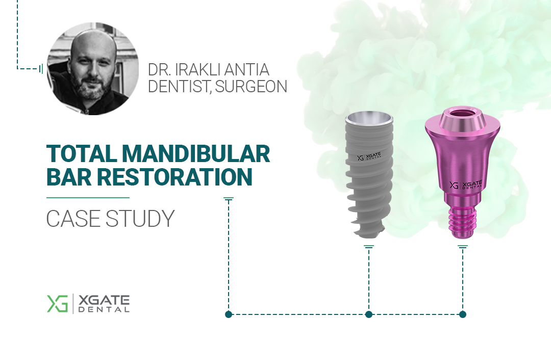
Let’s introduce the clinician who treated this patient:

Dr. Irakli Antia
Dentist, Surgeon

Ketevan Kushitashvili
Prosthodontist
Results of the initial examination of the patient
The patient was a woman in her 70s with a stable general health condition and controlled blood pressure. No acute conditions present.
Her primary complaint was poor retention of her existing removable lower denture.
A Cone Beam Computed Tomography (CBCT) scan, performed on October 17, 2024, revealed significant bone loss (both height and volume) in the lateral sections of the mandible. Only the anterior region was deemed suitable for implant placement.
The bone density was low, making immediate loading impossible due to the lack of sufficient primary stability. A two-stage protocol was therefore selected: first, implant placement with plugs, and then, after osseointegration, placement of a temporary bridge, followed by a final prosthesis supported by a bar.
The bar was chosen as the support structure to evenly distribute chewing forces across the four implants and compensate for the cantilever forces generated by the distal extensions of the restoration.
Selection of implants, abutments and implementation of the treatment plan
Two primary factors influenced the implant diameter and length selection:
- Available bone height and width to adequately support the implants.
- The significant mechanical load that would be transmitted through the bar to the implants.
Therefore, implants with a 5 mm diameter and the maximum permissible length for the available bone volume were selected.
Implants were planned for the following positions:
- #32 – XGate X11 implant, 5 mm diameter, 11.5 mm length, 11° conical interface.
- #34 – XGate X11 implant, 5 mm diameter, 11.5 mm length, 11° conical interface.
- #43 – XGate X11 implant, 5 mm diameter, 11.5 mm length, 11° conical interface.
- #45 – XGate X11 implant, 5 mm diameter, 13 mm length, 11° conical interface.

XGate X11 implant, 5 mm diameter,
11.5 mm length, 11° conical interface

XGate X11 implant, 5 mm diameter,
13 mm length, 11° conical interface
The implants were placed on October 24, 2024. Plugs were placed, and the soft tissues were sutured.
One month post-surgery, after resolution of swelling and complete soft tissue healing, a removable temporary denture, passively retained to the gums, was fabricated.
The patient wore this prosthesis until mid-March 2025.
On March 11, 2025, the soft tissues were re-opened, and multi-unit abutments (MUAs) were placed on the implants. These acted as healing caps until March 29th to allow proper soft tissue healing and emergence profile development.

The choice of multi-unit abutments deserves special attention. All 4 implants were fitted with low-profile multi-units V-type by XGate, an example is in the following picture. Note that MUA V-type are available with different gingival heights – from 0.5 to 5 mm.


Based on the fact that one of the implants was placed slightly deeper than the others, the doctor chose the following modifications of multi-unit abutments:
#32 – MUA V-type – 2 mm
#34 – MUA V-type – 3 mm (due to the implant placed slightly deeper than the others)
#43 – MUA V-type – 2 mm
#45 – MUA V-type – 2 mm
Soft tissue healing around the abutments went without complications, as evidenced in the photo below.

A significant advantage of screw-retained restorations using MUAs is that the abutments remain in place throughout the restoration’s lifespan. This allows for uninterrupted epithelial and connective tissue attachment and ensures a hermetic seal between the implant and oral environment, minimizing marginal bone loss. Removing the healing caps and placing abutments will disrupt this formation during the try-in and adjustments of the prosthesis.
After soft tissue maturation, the patient received a temporary PMMA (polymethyl methacrylate) bridge on March 29, 2025.
On May 2, 2025, the patient received the final restoration: a titanium bar framework supporting a zirconium dioxide bridge.
The titanium bar that supports the entire restoration, including the distal crowns, is shown in the image below.


The table below summarizes the key aspects of this clinical case, including details of the implants and abutments used.
| Treatment stage | What was done | Notes | |||||||||||||
|---|---|---|---|---|---|---|---|---|---|---|---|---|---|---|---|
| Initial Examination and Diagnostics | Examination and CBCT scan. Results: Low bone density and insufficient bone volume in the lateral sections for implant placement. | Patient presented with complaints of poor lower denture retention. | |||||||||||||
| Treatment Plan | Two-stage protocol: Placement of 4 implants in the anterior mandible, followed by a zirconia restoration supported by a titanium bar after complete osseointegration. | A two-stage protocol was chosen due to low bone density. Support on 4 implants was necessitated by insufficient bone volume in the posterior regions. | |||||||||||||
| Implantation | 4 implants successfully placed on 10/24/2024. | XGate Dental implants with an 11° conical interface were selected to ensure secure retention of the platform‐switching abutments. | |||||||||||||
| |||||||||||||||
| Fabrication of Temporary Denture | Polymer removable denture retained by the gingiva. | Placed one month post-implantation. | |||||||||||||
| Multi-Unit Abutment Placement | Soft tissues exposed, and 4 low-profile (1.5 mm) multi-unit abutments placed on 03/11/2025. | One multi-unit abutment was longer to compensate for the deeper implant placement at the #34 position. | |||||||||||||
| |||||||||||||||
| Temporary Screw-Retained Prosthesis Placement | A temporary PMMA bridge was manufactured and successfully placed on 29/03/2025. | Time was allowed between MUA placement and temporary bridge insertion for soft tissue healing and development of the emergence profiles. | |||||||||||||
| Final Restoration Placement | A titanium bar framework was placed with a zirconium dioxide prosthesis. | The entire treatment duration, from initial examination to final prosthesis placement, was approximately six months. Considering the patient’s age and bone condition, this represents an excellent outcome. |
Download Resources
Get the complete case study and explore our product catalog
Case Study • PDF • 1.3 MB
📥 Download Case StudyProduct Catalog • Online
🔍 View Product CatalogE-mail: [email protected]
Phone: (302) 498-8373
350 W Passaic
St Rochelle Park, NJ 07662
United States
XGate Dental Group GmbH
Falkensteiner Straße 77, 60322
Frankfurt am Main
Germany
Disclaimer: Any medical or scientific information provided in connection with the content presented here makes no claim to completeness and the topicality, accuracy and balance of such information provided is not guaranteed. The information provided by XGATE Dental Group GmbH does not constitute medical advice or recommendation and is in no way a substitute for professional advice from a physician, dentist or other healthcare professional and must not be used as a basis for diagnosis or for selecting, starting, changing or stopping medical treatment.
Physicians, dentists and other healthcare professionals are solely responsible for the individual medical assessment of each case and for their medical decisions, selection and application of diagnostic methods, medical protocols, treatments and products.
XGATE Dental Group GmbH does not accept any liability for any inconvenience or damage resulting from the use of the content and information presented here. Products or treatments shown may not be available in all countries and different information may apply in different countries. For country-specific information please refer to our customer service or a distributor or partner of XGATE Dental Group GmbH in your region.
