Contents
Almost every implantologist sooner or later experiences a patient’s wince and feels an implant spin during prosthetic procedures. This most often occurs in the maxilla during abutment placement. The most important thing in this situation is to remain calm and avoid any sudden movements. Mechanical failure of osseointegration does happen, and it is not always the clinician’s fault. The insertion torque may have been within the normal range, the bone quality may have appeared adequate, and the healing time may have been sufficient, yet the implant failed to achieve acceptable secondary stability. When the clinician applies the requisite 35 Ncm of torque to seat the abutment, the implant spins. This is a relatively rare but solvable problem. In this article, we will explore the available literature on this topic and outline the immediate steps a clinician should take.
A Study Confirming the Possibility of Reosseointegration
It is important to note that removing the implant is often unnecessary; in most cases, reosseointegration will occur within two months, allowing prosthetic procedures to resume. Our goal is to provide a theoretical basis for this recommendation. While there are few publications on this topic, we found one high-quality study published in 2013 by researchers from Korea: Reosseointegration of mechanically disintegrated implants in dogs: mechanical and histometric analyses.
The studies were conducted on beagle dogs, with implants placed on both sides of the mandible. To simulate a lack of primary stability, the implants were intentionally placed with virtually zero insertion torque, meaning the diameter of the final drill was equal to the implant diameter. This contrasts with standard surgical protocols, where the osteotomy is slightly undersized to ensure the implant threads engage and compress the surrounding bone to achieve primary stability. This method was used in both the control and experimental groups.
After four weeks, the soft tissues in the experimental group were reflected, the cover screws were removed, and resonance frequency analysis (RFA) was performed to determine the Implant Stability Quotient (ISQ). Despite the suboptimal placement protocol, the implants had already achieved significant integration by this point. After the measurement, the implants were mechanically reversed out, photographed, and re-seated into the same sites.

Retrieved implant with tissue remnants during a reosseointegration test (to be returned to the original site). Youtube/ Implantarium/ Rauf Aliyev
Upon re-insertion, the implants were only hand-tightened, as documented in the study protocol.
Immediately after the implants were returned to their sites, the ISQ was measured again.
The cover screws were then replaced, and the soft tissues were sutured. After another four weeks, all implants in both the control and experimental groups were uncovered.
At this stage, a full examination was conducted. Stability was measured again using RFA and the Periotest device. This was followed by a complete histological examination, which provides the most interesting insights. The image below shows a histological section from the control group, where the implants remained undisturbed for the entire 8 weeks.

Histological section of tissue at the implant interface (control group): black arrows indicate the margin of the osteotomy. Youtube/ Implantarium/ Rauf Aliyev
In the magnified image (500-micron scale), the black arrows indicate the margin of the original osteotomy. It is also clear that the osteotomy was wider than the implant threads. In the space between the osteotomy margin and the implant surface, well-formed bone tissue is visible. It is worth noting that metabolic processes are faster in dogs; in humans, the tissue would not be as mature after 8 weeks.
The tissue sections from the experimental group also showed excellent osseointegration.

Histological section of tissue at the implant interface (experimental group): the healing process is at an earlier stage compared to the control group, but signs of normal osseointegration are present. Youtube/ Implantarium/ Rauf Aliyev
Naturally, there is no clear boundary between old and new bone in this group due to the significant trauma caused by implant removal. However, the bone tissue near the implant surface is of acceptable quality, although there are visible areas of coarse, fibrous (woven) bone not seen in the control group—a sign of incompletely remodeled bone tissue. Overall, a normal healing process with comparable results was observed in both groups.
In comparing the enlarged images, the control group exhibits more mature lamellar bone, while the experimental group shows a higher proportion of fibrous bone. Nonetheless, in both cases, normal healing and osseointegration occurred.

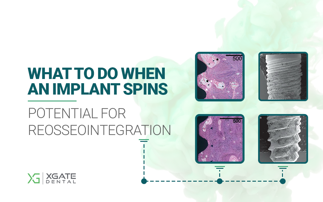
Comparison of histological sections from the control (left) and experimental (right) groups. Both show signs of normal healing, though the experimental group shows a slight lag in maturation. Youtube/ Implantarium/ Rauf Aliyev
Another interesting observation was made in the experimental group. High magnification revealed that new bone tissue had integrated with fragments of previously formed bone. These fragments remained on the implant surface during its retrieval at 4 weeks. The image of the removed implant clearly shows tissue remnants in the thread recesses. These bone fragments, already integrated with the titanium, served as nucleation sites for new bone formation.

Representative micrographs of the experimental group: High magnification shows the formation of woven bone around pre-existing bone fragments on the implant surface and in the native bone bed. Appositional bone growth was observed on the exposed implant surface, extending from these residual bone fragments. YouTube/ Implantarium/ Rauf Aliyev
The results of this study confirm that reosseointegration of implants is possible, providing documented evidence for what experienced clinicians have long advised in cases of stability failure: there is no need to immediately remove and replace the implant. We will later describe the precise protocol for managing a mobile implant with a partially seated abutment.
Which Method of Implant Stability Testing Performed Best?
During the study, implant stability was tested using two methods:
- Resonance Frequency Analysis (RFA) to determine the Implant Stability Quotient (ISQ).
- The Periotest device, which uses an impact-based mechanical method (electronic percussion).
Non-contact RFA provided relatively clear and consistent results in measuring the actual stability of the implants. Other studies also confirm that ISQ is currently the most reliable clinical method for determining implant stability, short of histological analysis.
The Periotest results showed a wide scatter of values, from which no definitive conclusions could be drawn.
Results of ISQ Measurements in the Control and Experimental Groups
In the experimental group, the average ISQ reached 77 by week 4. A repeat measurement immediately after removal and repositioning showed a decrease in the ISQ to 70. This highlights a limitation of RFA; a freshly removed and hand-tightened implant has virtually zero mechanical stability, yet the device indicates a value that might be considered sufficient for immediate loading. However, barring such exceptional circumstances, the ISQ generally provides a realistic assessment, as confirmed by numerous studies.
The most interesting results were obtained eight weeks after the start of the experiment. The average ISQ score was significantly higher in the experimental group compared to the control group.
- Control group average ISQ = 68.80 ± 5.02
- Experimental group average ISQ = 73.80 ± 5.85
And this is not a coincidence, its reliability is confirmed by the level of p = 0.042.
It may seem counterintuitive, but after the traumatic event, not only was osseointegration restored, but the implant became more stable.
Let’s examine this in more detail. Electron microscope images clearly show fragments of integrated bone tissue distributed across the entire surface that was embedded in bone.

Electron microscope images of implants retrieved after 4 weeks: (a) coronal view; (b) apical view. Residual bone tissue integrated into the surface is found across the entire implant. YouTube/ Implantarium/ Rauf Aliyev
Furthermore, histometric analysis of the bone-to-implant contact (BIC) showed that the average BIC value across all areas of micro- and macro-threads was higher in the experimental group (p = 0.043).
The conclusion published by the Korean researchers was:
“Within the limitations of this animal study, it can be concluded that an implant can be successfully reosseointegrated by unloaded submerged healing for a certain period of time following the mechanical breakage of osseointegration.”
Advantages and Disadvantages of Resonance Frequency Analysis of Implant Stability
On the one hand, RFA provides reliable indicators that reflect clinical reality. On the other, it “failed” when a mobile, hand-tightened implant showed only a slight decrease in ISQ.
The reason lies in the technique’s principle of operation. RFA detected that the implant surface was still connected to a significant amount of bone tissue. This means that over a large portion of the surface, the bond between the titanium and the initial layer of bone was stronger than the cohesive strength of the newly formed bone itself. This is why numerous bone fragments remained on the surface of the retrieved implant. The device registers this connected bone mass and cannot detect the micro-fracture plane within the bone.
While it is difficult to imagine this scenario in clinical practice—no one would proceed with prosthetics on an implant that was just spun—it does suggest that 6-8 weeks after a mechanical stability failure, prosthetic treatment is indeed feasible.
Clinical Example: Confirmation of Successful Reosseointegration
The initial conditions for implantation were virtually perfect, with ample bone volume and sufficient soft tissue. The treatment plan was to place three implants in the first quadrant to support a screw-retained, three-unit fixed partial denture.
The only concern was the risk of implant fenestration at site #14. Here, the alveolar ridge had a “pear-shaped” profile with sufficient bone coronally but a constriction at the apex, requiring precise implant positioning.

Potential for implant fenestration at site #14 due to alveolar ridge anatomy. Youtube/ Implantarium/ Rauf Aliyev
This is a manageable problem. A flap was raised, the periosteum was elevated, and an allograft-based bone graft was placed over the defect. This is a standard Steigmann technique. No membrane was required; the bone graft was contained by the periosteal flap, which was secured with a periosteal suture.

Bone defect closure using the Steigmann technique (periosteal envelope). Youtube/ Implantarium/ Rauf Aliyev
Next, healing caps were placed, and the soft tissue was sutured. The patient was dismissed for a three-month healing period. The images below show the site immediately after surgery and three months later. Healing was uneventful, and the gingiva appeared healthy.

Implants with healing caps: immediately after surgery (left) and three months later (right) Youtube/ Implantarium/ Rauf Aliyev
Percussion testing went well; all implants were immobile and produced a sharp, clear sound. The radiograph also revealed no issues.
Everything appeared to be perfect: the gingiva was in excellent condition, with no signs of fistulas or inflammation. However, the implant at site #17 spun during the torquing of the multi-unit abutment. The implants at sites #14 and #16 remained stable.
Based on clinical experience and the principles outlined in the study above, the clinician decided not to remove the implant. The multi-unit abutment that caused the failure was left in place, a healing cap was placed over it, and a shortened two-unit bridge was fabricated for the successfully integrated implants. The ability to leave the multi-unit abutment in place was advantageous; if a cement-retained restoration had been planned, attempting to unscrew the abutment would have likely resulted in the complete removal of the implant.

A healing cap was placed over the MUA on the mobile implant, and a shortened bridge was placed on the two stable implants. The compromised implant was left alone for the time required for reosseointegration. Youtube/ Implantarium/ Rauf Aliyev
The patient received the partial restoration and was scheduled for a follow-up in two months to allow for reosseointegration. However, as often happens, the patient returned late, after six months. This was in June 2020.
Radiograph and clinical view after a 6-month break. Youtube/ Implantarium/ Rauf Aliyev
Unfortunately, the clinician did not have an ISQ device at the time, so percussion testing was performed again through the healing cap. The sound was sharp and clear. The implant passed a preliminary test when the clinician removed the multi-unit abutment; the implant remained stable.
The bridge was temporarily removed; the gingival condition around all implants was excellent. The decision was made to proceed with a single, screw-retained restoration for the third implant and to remake the bridge to include all three units.

Checking the condition of soft tissues before installing a bridge and single-tooth restoration. YouTube/ Implantarium/ Rauf Aliyev
After removing the temporary bridge, a scan body was placed on the final implant. Based on the digital impression, a single zirconia crown was fabricated on a tall titanium base. The single-unit restoration was also torqued to 35 Ncm, which served as the final confirmation of its stability.
The images below show the final three-unit restoration as of September 2020.

Final restoration: bridge and single restoration installed in September 2020. Youtube/ Implantarium/ Rauf Aliyev
The patient returned for a follow-up examination one year later, in September 2021. The gingiva was healthy, with no signs of recession or inflammation.

A photo of the restoration one year after the final version was installed – a bridge + single crown: the soft tissues are healthy. YouTube/ Implantarium/ Rauf Aliyev
The follow-up radiograph also showed that everything was all right. The bone tissue around the implants was even in better condition after a year.

Control radiograph 12 months after restoring the previously mobile implant. YouTube/ Implantarium/ Rauf Aliyev
This is a standard outcome in such situations. In the absence of complications like inflammation or radiographic signs of pathology, the implant fully re-integrates and can provide service for decades.
How to Eliminate the Risk of Mechanical Disintegration of the Implant?
Currently, the most reliable method is to use RFA to measure the ISQ before initiating prosthetic treatment. Many experienced prosthodontists recommend not proceeding with prosthetics if the ISQ value is below 70, even though the official scale considers a value of 65 sufficient.
While an ISQ device may not be necessary for every case, it is an invaluable tool in situations where implant stability is uncertain.
That’s all for now, stay tuned for the next publication.
Disclaimer: Any medical or scientific information provided in connection with the content presented here makes no claim to completeness and the topicality, accuracy and balance of such information provided is not guaranteed. The information provided by XGATE Dental Group GmbH does not constitute medical advice or recommendation and is in no way a substitute for professional advice from a physician, dentist or other healthcare professional and must not be used as a basis for diagnosis or for selecting, starting, changing or stopping medical treatment.
Physicians, dentists and other healthcare professionals are solely responsible for the individual medical assessment of each case and for their medical decisions, selection and application of diagnostic methods, medical protocols, treatments and products.
XGATE Dental Group GmbH does not accept any liability for any inconvenience or damage resulting from the use of the content and information presented here. Products or treatments shown may not be available in all countries and different information may apply in different countries. For country-specific information please refer to our customer service or a distributor or partner of XGATE Dental Group GmbH in your region.

