Contents
Before we discuss the study’s design and results, let’s briefly review the range of multi-unit abutments available for Full Arch-on-4/6 restorations. XGate Dental has developed three types of multi-unit abutments designed for immediate loading protocols like Full Arch-on-4/6:
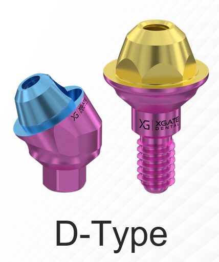
- MUA D-type. This is the classic and earliest version in the line, available in both straight and angled options.
 This is a proven and effective option. The 17° and 30° angled versions are particularly noteworthy, as they can compensate for a total implant divergence of up to 100°.
 These abutments also feature convenient color-coding to indicate gingival height.
A typical Full Arch-on-4 prosthetic solution is illustrated below.
- MUA V-type. These are universal abutments with a reduced profile and an increased contact area between the abutment platform and the sleeve compared to the MUA D-type.
 The increased contact area of 10 mm² enhances the stability and overall mechanical strength of the structure.
This series only includes straight abutments, but the MUA V-type can accommodate moderate inter-implant angulations of up to 40°, which is often sufficient for Full Arch-on-4/6 prosthetics.
These multi-unit abutments are also conveniently color-coded by gingival height.
- MUA S-type. This is the latest development in abutments, featuring super-compact sizes for screw retention, which is especially valuable in aesthetic prosthetics. These multi-unit abutments are also color-coded by gingival height.
 A specially designed sleeve for the MUA S-type allows for angled insertion of the retaining screw. This is particularly relevant in the anterior region, where the screw access channel can emerge on the facial surface of the crown, creating an aesthetic issue. The screw channel is typically filled with composite, but over time, this composite can stain and discolor, becoming noticeable against the crown. By angling the screw channel, it can be repositioned to a less visible area, improving the final aesthetic outcome. The sleeve with an angled screw channel was developed specifically for this purpose.
The illustration below shows a comparison of the exact dimensions of the different types of multi-unit abutments from XGATE Dental.
XGATE Dental manufactures implants and all types of abutments from Grade 23 titanium alloy. However, we are seeing an increasing number of publications about implants made from zirconia, other advanced ceramics, and even high-performance polymers. Therefore, we decided to share the results of an insightful study with our readers and discuss why laboratory findings don’t always translate directly to clinical practice.
This article is based on the FEA study “Mechanical Integrity of All-on-Four Dental Implant Systems: Finite Element Simulation of Material Properties of Zirconia, Titanium, and PEEK”
In daily implantology practice, discussions often revolve around implant shape, length, and position. However, one of the most critical factors—the material of the implant system—is often overlooked. Yet, it is the material that ultimately determines how the restoration will behave under occlusal loading after one, five, or ten years.
The study we are reviewing today focuses on three materials that are increasingly part of the clinical choice:
- Titanium—The “workhorse and gold standard” of implantology.
- Zirconia—Aesthetic, strong, and rapidly gaining popularity.
- PEEK—A lightweight polymer that offers biomimetic properties.
Researchers conducted a comprehensive finite element analysis (FEA), modeling an Full Arch-on-4 prosthesis under realistic load conditions. This type of simulation allows us to visualize what is impossible to assess with the naked eye: how stress is distributed within the implant, where deformation occurs, and where the risk of stress concentration is highest.
The implant architecture in the model was complex, including three fences (support ribs) to simulate real structural elements at the bone-implant interface.
The study’s findings were rich and, in some respects, unexpected. We have prepared this review to summarize the research for dental surgeons and general practitioners who need to understand the mechanical properties of materials, not just their aesthetic appeal.
How the Study Was Conducted: A Brief Overview
FEA model
The researchers created a 3D model of the implant in CATIA and performed the simulation in ANSYS:
- implant length:8 mm
- diameter:4.5 mm
- three “fences”: two 2 mm, one 1.5 mm
- bone: 1 mm cortical layer + trabecular bone
- 81,000 nodes, 27,000 mesh elements
The model was the same for all materials to eliminate the influence of geometry.
Materials
The study compared:
- titanium
- zirconia
- PEEK (polymer)
All components (bone and implant) were modeled as linear elastic and isotropic, which allows for a valid comparison of material properties.
Loads
Two modes:
- Perpendicular load 100 N—for each of the four ends → total 400 N.
- Oblique load 100 N at an angle of 30°—simulation of the trajectory of chewing forces.
Three key metrics were measured:
- deformation (µm)
- stress (MPa, von Mises)
- strain
Properties of Materials: The Foundation That Explains Almost Everything
The table below shows the mechanical characteristics that determine the behavior of the implant.
Table 1. Mechanical Properties of Materials
| Material | Young’s modulus (GPa) | Tensile strength (MPa) | Density | Features |
|---|---|---|---|---|
| Zirconia | 210 | 1000–2000 | 6,05 | Very hard, durable, low ductility |
| Titanium | 96 | ~930 | 4,62 | Good combination of strength and elasticity |
| PEEK | 4 | 150–215 | 1,31 | Soft, flexible, modulus close to bone |
Why is this important?
- The higher the Young’s modulus → the less the material deforms, but the more concentrated the stress becomes.
- The softer the material → the more it deforms, but creates fewer peak stresses.
This explains why zirconia deforms little, while PEEK deforms a lot.
Results: Perpendicular Load
Deformation
Table 2. Deformation Under a Load of 100 N (Perpendicular)
| Material | Max. deformation (µm) | Average deformation (µm) | Note |
|---|---|---|---|
| Zirconia | 1.267 µm | 0.198 µm | ↓84.91% relative to PEEK |
| Titanium | 1.745 µm | 1.459 µm | Similar max. to zirconia, but higher average |
| PEEK | 8.404 µm | 5.598 µm | The highest deformation |
Key takeaway: Zirconia is the most rigid material, showing the least deformation in both maximum and average values.
Stress
Table 3. Maximum and Average Stress (MPa)
| Material | Max. stress (MPa) | Average stress (MPa) | Dispersion rate (%) |
|---|---|---|---|
| Zirconia | 15.477 | 0.461 | 97.0% |
| Titanium | 13.507 | 4.503 | 66.7% |
| PEEK | 8.967 | 4.553 | 49.2% |
How to interpret the “dispersion rate” column?
This is the ratio of average to maximum stress: the closer it is to 100%, the more evenly the load is distributed.
And here zirconia shows a result that stands out in the study.
Deformation vs. Stress: What Matters More in Practice?
- Zirconia is almost non-deformable and dissipates loads perfectly..
- Titan—compromise between flexibility and strength.
- PEEK—soft material, which reduces peak stresses, but undergoes high deformation.
Results: Angled Load (Chewing Trajectory)
When loading at an angle, the differences between the materials become even more noticeable.
Table 4. Deformation Under a Load of 100 N at an Angle of 30°
| Material | Max. deformation (mm) | Average (µm) |
|---|---|---|
| Zirconia | 0.208 mm | 14.43 µm |
| Titanium | 0.277 mm | 18.49 µm |
| PEEK | 0.939 mm | 45.89 µm |
What is important:
- PEEK deforms by an order of magnitude more than the other materials.
- Zirconia once again demonstrates the most stable geometry.
Table 5. Stress (Oblique Load)
| Material | Max. stress (MPa) | Average stress (MPa) | Dispersion (%) |
|---|---|---|---|
| Zirconia | 611.6 | 5.72 | 99.06% |
| Titanium | 422.6 | 5.05 | 98.80% |
| PEEK | 249.99 | 4.50 | 98.19% |
Key takeaway:
Despite the high maximum stress of zirconia, its average stress remains the lowest, which indicates an even distribution of the load.
Key Metrics Comparison Chart (Mermaid)
Discussion: What Does This All Mean for Clinical Practice?
Zirconia: Minimal Deformation + Better Load Distribution
The study shows that zirconia acts as a rigid framework.
It absorbs the load and barely changes shape. This allows the load to be distributed more evenly throughout the entire structure, including the fences.
Because of this, zirconia may be particularly interesting for Full Arch-on-Four, where each implant carries a significant share of the load.
Titanium: The Golden Mean
Titanium behaves predictably:
- Moderate deformation.
- Acceptable stress distribution.
- Stress concentrations are lower than zirconia’s but higher than PEEK’s.
It is this combination of properties that has made titanium the industry standard.
PEEK: low stress but high deformation
The results showed:
- It reduces peak stress (a potential advantage).
- However, it deforms significantly (a potential risk).
High deformation at the implant level leads to:
- Load shifting
- Stress concentrations within the structure
- Potential points of fatigue failure
In the study, the authors note that such a model of deformation can accelerate mechanical complications, especially in long-span restorations.
What Doctors Should Remember: Key Interpretations
(No recommendations – only interpretation of study results.)
- Zirconia in this model showed
- minimal deformation
- minimal average stress
- the most even distribution of the load.
- Titanium demonstrated a stable average performance, falling between the other two materials on most parameters.
- PEEK—a material that reduces peak loads, but loses out in terms of deformation and stress distribution.
- Under oblique loading, the differences between the materials become even more striking,
- Full Arch-on-4 is a system in which every implant is critical.
Therefore, material properties play a particularly important role.
Summary Table of Key Metrics
Table 6. Comparison of Materials by Key Mechanical Characteristics
| Metrics | Titanium | Zirconia | PEEK |
|---|---|---|---|
| Deformation (perpendicular) | 1.745 µm | 1.267 µm | 8.404 µm |
| Average deformation | 1.459 µm | 0.198 µm | 5.598 µm |
| Max. stress (perpendicular) | 13.5 MPa | 15.5 MPa | 8.97 MPa |
| Moderate stress | 4.50 MPa | 0.46 MPa | 4.55 MPa |
| Load dissipation | 66.7% | 97.0% | 49.2% |
| Deformation at an angle | 0.277 mm | 0.208 mm | 0.939 mm |
| Stress at an angle (medium) | 5.05 MPa | 5.72 MPa | 4.50 MPa |
Conclusion
During the FEA analysis of three materials in the Full Arch-on-4 configuration, the researchers obtained an interesting and largely unexpected set of results:
- Zirconia demonstrates better mechanical stability with minimal deformation.
- Titanium maintains its position as a universal material, providing balanced properties.
- PEEK has low peak stresses, but is significantly inferior in terms of deformation.
The study emphasizes the importance of choosing a material as an independent factor in the long-term durability of a restoration.
Editorial Comments
A FEA study, even a well-conducted one, cannot replace extensive clinical practice and long-term observation.
In short, the essence of the contradiction
- The FEA model showed that zirconia in this geometry “wins” by two criteria: Minimal deformation and very high “dispersion” of the average stress (in the model, the load is distributed uniformly throughout the structure). This makes zirconia “rigid and stable” under the simulated conditions.
- Clinical practice and fatigue testing point to another limitation of zirconia: compared to titanium it is more fragile (less plasticity, lower impact strength), therefore, under real-world conditions (especially in case of cyclic chewing load, seating errors, thin cross-sections, misalignment), it can crack or experience “mechanical” failures more often than titanium.
In other words, FEA examines one dimension—stress distribution/deformation under ideal conditions.The clinical reality involves fatigue, microcracks, connections, material defects and many years of operation.
Expanded Analysis: Pros and Cons of Each Material
Zirconia – Pros (FEA and Material Data)
- Very high modulus of elasticity → minimal deformation under load (FEA results).
- Good ability to “dissipate” average stresses in models, with proper design – low average stress.
- Aesthetics: no “gray shine”, useful in the frontal area.
- High resistance to wear and corrosion (ceramics).
Zirconia – Cons (Clinical Risks)
- Fragility and risk of cracking: Zirconia is a ceramic, it has limited ductility; fatigue cracks occur at local stress concentrators.
- Features of the “implant-abutment” connection: Two-piece zirconia implants/abutments require very careful design of the connection; poor connection design leads to a high risk of microcracks and fractures.
- Limited long-term clinical data for two-piece zirconia systems (especially >5–10 years) compared to titanium.
- Sensitivity to manufacturing defects (pores, thermal stresses during sintering).
Titanium (Grade 5, Grade 23) – Pros
- High viscosity and plasticity: The metal bends, but does not break suddenly; this gives it a “safety margin” against overloads and fatigue.
- Excellent fatigue strength: Titanium and its alloys (Grade 5 – Ti-6Al-4V; Grade 23 – ELI variant) and decades of proven clinical reliability.
- Design of connections (screw connections, platform compatibility): Extensive testing, many clinical protocols.
- Forgiveness in case of minor discrepancies and installation errors.
Titanium – Cons
- Aesthetics: A bluish tint may show through in case of a thin gingiva.
- Corrosion under rare adverse conditions (reaction with the environment), but this issue is generally clinically manageable.
PEEK: Pros and Cons (Briefly)
- Pros: Modulus of elasticity is closer to bone, good for temporary restorations, absorbs shock, reduces peak stress.
- Cons: Low rigidity leads to high deformation; its behavior under long-term cyclic loading, especially in thin sections, raises questions. Currently, its primary role is for specific applications (e.g., temporary superstructures, shock-absorbing components) rather than as a universal replacement for titanium.
What the Totality of Evidence Suggests (Common Findings)
- Titanium remains the “gold standard” due to its long-term clinical reliability, predictability, and resistance to fatigue. This is not a marketing statement but the result of decades of clinical observation and meta-analyses.
- Zirconia is attractive because it deforms less in models and offers superior aesthetics. However, its brittleness and tendency to fracture under real-world loads are the reasons why clinicians approach it with caution for full-arch, implant-supported restorations.
- Two-piece zirconia systems (implant + zirconia abutment) have historically shown a higher rate of technical complications compared to titanium systems if the design and connection are not perfectly executed.
- FEA is a useful tool, but it does not replace clinical trials. It models stress in an idealized environment, whereas long-term fatigue strength is influenced by real-world factors like micro-defects, component fit, and dynamic patient forces.
We hope this article was useful for you. Stay tuned for our next publication.
Disclaimer: Any medical or scientific information provided in connection with the content presented here makes no claim to completeness and the topicality, accuracy and balance of such information provided is not guaranteed. The information provided by XGATE Dental Group GmbH does not constitute medical advice or recommendation and is in no way a substitute for professional advice from a physician, dentist or other healthcare professional and must not be used as a basis for diagnosis or for selecting, starting, changing or stopping medical treatment.
Physicians, dentists and other healthcare professionals are solely responsible for the individual medical assessment of each case and for their medical decisions, selection and application of diagnostic methods, medical protocols, treatments and products.
XGATE Dental Group GmbH does not accept any liability for any inconvenience or damage resulting from the use of the content and information presented here. Products or treatments shown may not be available in all countries and different information may apply in different countries. For country-specific information please refer to our customer service or a distributor or partner of XGATE Dental Group GmbH in your region.

