For those unfamiliar with XGate Dental products, it may not be immediately apparent how V-type multi-unit abutments can address specific challenges in dental practice. This article presents several clinical cases demonstrating the application of MUA V-type solutions, valuable for both novice and experienced practitioners.
What are Multi-Unit Abutments V-Type?
Let us recall the main advantage of screw retention. When the vertical space from the implant platform to the opposing tooth is less than 7 mm, screw retention is often the preferred method. Cement retention is generally not recommended in these situations due to the risk of cement failure and difficulty in cleaning excess cement.
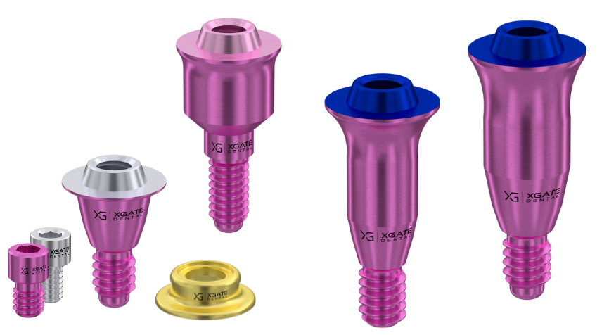
All multi-unit abutments are designed to facilitate screw retention for multiple-unit restorations. Standard D-type multi-unit abutments typically have an interface height of 3.5 mm, a dimension consistent among various manufacturers. However, many clinical situations require a lower profile. XGate has developed an ultra-low V-type multi-unit abutment, with a height of only 1.5 mm. Several MUA V-type options are available to accommodate diverse clinical scenarios, with variations in gingival height and abutment body shapes (see illustration below).

Varieties of multi-unit abutments V-type by XGate
The design of these abutments enables platform switching and the creation of an optimal emergence profile in a variety of clinical contexts. Gingival heights range from 0.5 mm to 5 mm.
Special sleeves, available in different heights, are used to attach the restoration to the multi-unit abutment (see illustration below).

Sleeves for attaching the prosthesis to multi-unit abutments V-type
For example, a 4 mm sleeve is a standard option used in approximately 95% of cases. It provides sufficient contact area for cementation while remaining compact enough to accommodate a crown. A 3 mm sleeve is shorter, allowing for smaller crown heights, but it offers a reduced contact area for cementation. While a 3 mm sleeve can still provide adequate retention, the screw may exert some pressure on the restorative material itself, rather than solely on the sleeve, as with the 4 mm version.
The 1.5 mm sleeve is reserved for extreme cases. In this instance, the entire screw resides within the restorative material (e.g., zirconia or PFM bridge metal), and there’s minimal contact between the screw and sleeve. The 1.5 mm sleeve provides a minimal surface for cementation, necessitating meticulous cementation techniques and the use of a strong adhesive.
Six-millimeter sleeves are indicated for cases involving very tall crowns, ensuring that forces on the crown do not compromise the cement. However, this is a less common application.
Now, let’s examine specific cases where the utility of V-type abutments becomes evident in challenging clinical situations.
Case #1: 80-Year-Old Man with Complicated Implant Placement
The patient presented with a damaged and aesthetically compromised upper jaw restoration that had been in place for over 20 years. The existing screw-retained prosthesis had a metal framework veneered with composite resin, which had experienced wear and chipping, particularly in the area of the anterior incisors.
Compounding the issue, one of the implants placed two decades prior exhibited a significant buccal inclination, resulting in the screw access channel exiting on the facial surface of the tooth. The composite used to seal the channel had discolored.
The lower bridge was also in poor condition, despite lacking obvious defects.

Initial situation: damaged upper bridge with a pigmented spot on the composite covering the screw shaft; significant wear and loss of aesthetics on the lower bridge
The treatment plan involved fabricating new bridges from zirconia, utilizing the existing implants for support. The patient’s health status precluded the placement of additional implants or other surgical interventions. The patient, who was 80 years old and wheelchair-bound, primarily sought aesthetic improvements. He reported no functional limitations with the existing bridges.
The clinician faced a challenging task: designing and fabricating a new prosthesis while eliminating the undesirable facial screw access channels on both the upper and lower bridges.
The existing bridges were removed, one of which is shown below. The bridge exhibited considerable wear but maintained functionality.

A bridge that served for 20 years was removed
The original bridges had a metal framework. The new bridges were to be monolithic zirconia. This presented a challenge given the angulation of the implants in the maxilla. This is illustrated below.

Digital impression of the patient’s upper jaw made with an intraoral scanner
Metal frameworks can be cast to accommodate a variety of angulations and screw channel emergence profiles.
CAD/CAM milling of a prosthesis has limitations on the angulation between implants, typically no more than 30-40°.
If this were a different patient, an acceptable solution would be to place another implant next to the problematic one with the buccal inclination. Another option would be to not use the buccal inclined implant.
In this case, implant placement was not an option, and reducing the support from four to three implants was not an option either. The bridge was too long to be supported by three implants.
Another option would be to use an angled abutment, which would redirect the screw channel. But the angled abutment would be visible between the restoration and gingiva, which was unacceptable for aesthetic reasons.

Angled multi-unit abutment D-type: in this case the entire part where the marking is applied would be visible
The following solution was used:
- The old abutments were removed along with the bridges. It was decided to design and manufacture new bridges using CAD/CAM technologies. The old abutments were not compatible with this technology.
- V-type multi-unit abutments were selected for the new restoration. Due to the reduced inter-arch space, the zirconia bridges would be relatively thin, and incorporating standard multi-unit abutments could compromise the strength of the bridge.
- For three implants, multi-units with a gingival height of 1 mm were selected. For the problematic implant, a minimum height of 0.5 mm was selected.

- To address the facial screw access channel, it was decided to use the implant without screw retention. The smallest 1.5 mm sleeve was seated on the abutment but not retained with a screw. The other implants used standard sleeves with screw retention. The result can be seen in the images below.


Zirconium dioxide upper jaw bridge
As the images show, the axis of the new abutment is directed too high, above the incisal edge of the natural teeth.


3D model of the upper and lower jaw with installed MUA V-type
A protrusion was made in the form of an elongated crown inside which was the sleeve without the screw channel.

A finished zirconia dioxide upper jaw bridge with a protrusion opposite an irregularly positioned implant
The clinical result was acceptable. In everyday use, the protrusion is not noticeable, remaining behind the upper lip. This meant that the patient’s main wish to improve aesthetics was fulfilled.

Smile with daily use of new bridges, patient’s wishes fulfilled
- The lower bridge replacement was performed in a standard manner. The image below shows the lower bridge separately, and then how it looks in the patient’s oral cavity.


Lower jaw bridge ready for placement (top); upper and lower bridges in the patient’s mouth (bottom)
Most likely, new zirconia bridges will last for the rest of the patient’s life.
The support surface of the V-type multi-unit and its sleeves provide comparable stability and strength to standard abutments, despite their smaller size. The strength exceeds any forces that the patient can create.
Case #2: Critical Space Deficit Between Implants and Antagonist Teeth
This case is an example of a situation in which only screw retention can help, but even standard multi-unit abutments did not have adequate space. Low V-type abutments were selected.
Typically, cement retention is used in the anterior region, especially in the maxilla. Cement retention avoids screw channels which can be on the facial surface of the crowns. Facial screw channels are common because implants placed in the anterior region often have a buccal inclination.
The problems in this case were two-fold. The implants were placed with a palatal shift, and the prosthesis should be at the level of the natural teeth.

3D image of the upper jaw with implants placed: it is visible that both implants are displaced palatally
The second problem was that the distance from the lower teeth to the gingiva on the buccal side was only 3.5 mm. The distance between the base of the implants and the antagonist teeth is slightly larger. This means that there is very little space for the abutment and crowns.

3D image in lateral projection, where the small distance between the lower tooth and the gingiva of the upper jaw is visible
More precisely, the available abutment height in this case was limited to just 2.5 mm. While it might be technically possible to cement a crown onto a 2.5 mm abutment, early de-cementation would be highly likely due to the insufficient contact area between the abutment and the internal surface of the crown. Another significant concern was the difficulty of thoroughly removing cement remnants given the implant placement and gingival configuration. Furthermore, direct cement contact with the gingiva poses a high risk of inflammation. However, ensuring adequate retention and strength takes precedence over the risk of soft tissue irritation in this particular scenario. Therefore, screw retention was selected because it is less dependent on abutment height. This then led to considering the type of restoration, with two primary options:
- a PFM bridge is an acceptable option, but we have an aesthetic zone, and the metal frame can show through the composite or ceramic layer and give a grayish tint.
- A bridge made of monolithic zirconia is the preferred option without aesthetic flaws. That is why it was chosen.
So, the type of retention is determined, the material of the prosthesis is chosen. But many experienced doctors have already noticed that even for a zirconia bridge with screw retention, there is a critical lack of space. And if standard components are used, it is true, because:
- The minimum height of the subgingival part for most abutment manufacturers is 1.5-2mm.
- The minimum height of the sleeve, which rests on the abutment, is 4-4.5mm.
- The minimum height of the superstructure above the implant level is 1.5+4=5.5 mm, and only 3.5 mm is available in this case.
But XGate has V-type multi-units that are only 0.5 mm high, and combining such an abutment with a 3 mm sleeve would fit within the allowable distance.

Sleeve for MUA V-type
The general shape of the bridge and the cavity for the bushing with the screw access channels can be seen below.


Digitally designed zirconia bridge using the smallest multi-unit and small sleeve
Even on the digital model it is already clear that the thickness of the zirconia on the buccal side is more than sufficient to provide good aesthetics, although on the occlusal side we are left with only 0.5 mm of zirconia, which is not very good, but acceptable.
The last thing worth mentioning is the digital protocol that was used at all stages.
Case #3: Bar-Supported Restoration – Palatal Shift and Improved Fit
Here, there are five implants in the anterior maxilla. All of the implants have a buccal inclination and this is the problem of the case.

Implants with already installed MUA V-type on which the bar for a removable denture will be attached
We needed to position the bar as palatally as possible and as close to the gingival margin as possible. The solution was to utilize low-profile and short MUA V-type abutments from XGate, as illustrated in the image above. Let’s examine the features and benefits of this approach.
Consider the alternative. If the bar were simply attached to standard straight abutments, the aesthetics of the restoration would be compromised. Each abutment acts as a direct extension of the implant, causing the bar attachments to be directed buccally along the axial inclination of each implant. The resulting bar would be positioned too far labially, creating an excessive buccal prominence. Consequently, the removable denture would need to be fabricated with a thin labial flange, increasing the risk of metal framework visibility through the acrylic.
However, by using a smaller multi-unit abutment, the labial shift of the bar is minimized. As you can see in the image, the bar attachments are relatively low-profile, and the bar itself is shifted palatally during fabrication. The posts containing the screw access channels protrude labially, optimizing the overall aesthetic outcome.

A fixed bar on which the removable denture will rest
The aesthetic problem has been solved. The removable denture is attached, and no metal is visible through the plastic, see the final photo below.

Removable denture with bar retention
Let’s delve a little deeper into the design and differences between the various types of MUAs. All multi-unit abutments feature a flat platform with a conical protrusion that houses the screw access channel. The height of this conical protrusion in a standard D-type multi-unit abutment is typically 2.5 mm, although slight variations may exist between manufacturers. In contrast, the conical protrusion on the MUA V-type extends only 0.8 mm above the platform. Refer to the image below, where a D-type abutment has been substituted for a V-type on the patient’s jaw model, highlighting the significant difference in height

Comparison of the interface height of multi-unit abutments V-type and D-type on a jaw model
This was done for example purposes only; for the actual restoration, all V-type multi-units were used.
In addition to the height, the MUA V-type is smaller in diameter compared to the standard ones, namely 4.5 mm instead of 5 mm. This also allowed us to slightly reduce the dimensions of the beam in this case.
At the same time, the mechanical properties of the screw retention on the MUA V-type are comparable to the standard MUA D-type.
A logical question may arise: Why not always use a reduced multi-unit abutment if it appears to offer only advantages?
In fact, if there is no space shortage, then it is often more convenient to work with standard D-type multi-unit abutments. Both straight and angled abutments are available. The availability of different types of MUA significantly expands the possibilities for prosthetic solutions.
Case #4: Broken Temporary Bridge – Screw Channel Failure
In this situation, we have a cracked temporary prosthesis, clearly visible below. As the prosthesis is screw-retained, the task is not only to fabricate a replacement, but also to address the underlying cause of the original fracture.

Crack in temporary restoration – bridge needs to be replaced
After removing the damaged bridge, the doctor discovered that the structure was weakened by a long 4 mm sleeve and a screw channel above it in the area of the lateral incisor position. The crack appeared exactly where there was a cavity. The illustration below shows where the screw channel and the cavity for the sleeve are located on the old broken bridge.

Model of an old bridge with a long 4mm bushing and a screw channel, which weakened its structure
The decision was made to eliminate the screw channel and reduce the height of the bushing, see the illustration below. The thickness of the material and the strength of the bridge at this point increased significantly.

Model of a new bridge with increased thickness due to the elimination of the screw channel and reduction of the bushing height
This solution will have almost no effect on the retention strength of the entire bridge. The primary load borne by the multi-unit in the area of the lateral incisor is the vertical force generated during jaw closure. To withstand this load, it is sufficient for the prosthesis to simply rest on the multi-unit, without screw fixation, as illustrated in the image below.

New temporary bridge without screw channel in the area of the second incisor
In addition, in the new version of the bridge, the height of the crowns in the frontal area was slightly reduced, i.e., they were partially removed from the bite. This will further reduce local overload in the anterior section. And it was possible to remove part of the prosthesis from the bite due to the rejection of a long sleeve and a screw channel. The overall thickness of the plastic and the strength in this area have increased, which means that a slight decrease in thickness on the occlusal side will not affect the strength.
And this is confirmed by practice. The new temporary bridge did not break and served its term. Then, according to the same principle, a permanent prosthesis was made of zirconia. We all know that zirconia is a very strong and rigid material, but it does not tolerate bending and torsion loads at all and can break. And any weakening of the structure by internal holes increases the risk of prosthesis breakage.
But this patient’s zirconia bridge lasted at least a year and a half without any problems; the patient has not returned for further examinations.
Case #5: Removal of Old Bridge and Hopeless Teeth, Two-Stage Implant Placement
The original bridge, which was attached to ground-down teeth, needs to be replaced. Pigmentation is visible, and the periodontium condition is not the best. After removing the bridge, it was clear that the teeth were hopeless and needed to be removed.

The initial condition of the bridge
According to the treatment plan, the patient will have a new bridge with screw retention. The removal of teeth and installation of implants will be divided into stages, as the patient has contraindications to immediate implantation.
In the first stage, the patient will have a temporary bridge installed, supported by pins inserted into the remnants of the roots of natural teeth, as shown in the picture below.

Pins that hold the first temporary bridge
Then the pins and root remnants were also removed. But the temporary bridge on the pins should last 5-6 months, while the healing and full integration of the implants takes place. The first temporary bridge with support on the pins is made very massive so that it does not break, although the aesthetics suffers.

The first temporary bridge with pin support
Next comes the stage of opening the soft tissues, choosing and installing abutments, and replacing the temporary bridge. The new bridge will already have screw retention, the same as the permanent bridge. The next picture shows that one of the pins on which the first temporary bridge rested did not hold and was removed along with the prosthesis.

Soft tissue condition after removal of the first temporary bridge
Given the complex configuration and location of the implants, V-type multi-units were chosen. In the picture above, it is noticeable that the abutments have different colors; this reflects the varying gingival heights selected to accommodate the individual clinical needs.
Next, we prepare the upper jaw for scanning with an intraoral scanner. We install scanning markers from the level of multi-unit abutments to accurately capture their position and orientation, and create a digital impression of the jaw.

Preparing for scanning with an intraoral scanner
The result is a digital file like this, where scan bodies convey the exact location and angle of the implants.

Intraoral scan results
A new bridge is developed in a digital environment, the screw access channel and line of contact with the gingiva are designed.

Development of a prosthesis in a digital environment
In a virtual environment, it is easy to make different tissues transparent and deep structures visible. Also, thanks to libraries with 3D images of abutments, bushings and other components, it is easy to select the necessary part by trying it on a virtual image.
The next slide also shows the advantages of V-type abutments: even standard 4 mm bushings do not weaken the structure of the prosthesis, and all screw channels come out in more or less optimal positions.

Visualization of the location of the bushings fixing the prosthesis
As a result, we got this kind of restoration with screw retention.

Screw-retained prosthesis on V-type multi-units
The last photo shows how the second temporary prosthesis sits on screw retention. The result is more than acceptable. The next stage is to address the lower jaw.

The appearance of the screw-retained prosthesis is encouraging
That’s all, if you have any questions about choosing properties or application features of V-type multi-unit abutments, please contact us in any convenient way.
Disclaimer: Any medical or scientific information provided in connection with the content presented here makes no claim to completeness and the topicality, accuracy and balance of such information provided is not guaranteed. The information provided by XGATE Dental Group GmbH does not constitute medical advice or recommendation and is in no way a substitute for professional advice from a physician, dentist or other healthcare professional and must not be used as a basis for diagnosis or for selecting, starting, changing or stopping medical treatment.
Physicians, dentists and other healthcare professionals are solely responsible for the individual medical assessment of each case and for their medical decisions, selection and application of diagnostic methods, medical protocols, treatments and products.
XGATE Dental Group GmbH does not accept any liability for any inconvenience or damage resulting from the use of the content and information presented here. Products or treatments shown may not be available in all countries and different information may apply in different countries. For country-specific information please refer to our customer service or a distributor or partner of XGATE Dental Group GmbH in your region.
